HEALTH
5 Body Parts That Still Baffle Scientists

You might know something like the back of your hand, but do you know it as well as your vomeronasal organ? The human body may be mostly familiar territory, but much like the bottom of the ocean, it still isn’t completely charted. Even doctors and biologists haven’t sorted it all out, despite centuries of dissections, surgeries, scans, and microscopic examinations.
New imaging techniques have revealed structures that conventional tissue preparations concealed, while organs once dismissed as useless holdovers have turned out to perform important jobs. In other cases, scientists know that a body part matters but can’t observe it easily enough to explain exactly how it works.
Some of its mysteries are hiding in plain sight, while other puzzles are tucked deeper inside us. From a temporary organ that keeps a fetus alive to mysterious onion-shaped structures inside the immune system, these five body parts show how much there is still to learn.
The Sinuses
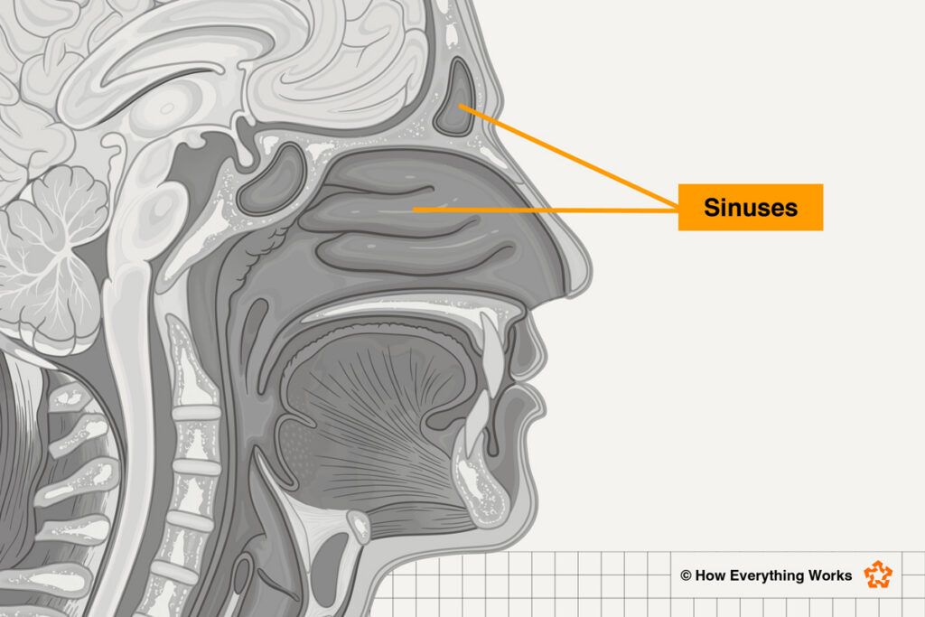
What are sinuses good for besides getting infected? Turns out, scientists aren’t exactly sure — especially when it comes to why we developed them in the first place. The paranasal sinuses (the ones most of us think of when we hear the word “sinuses”) are air-filled spaces tucked around the nose, eyes, and forehead. We know that their mucus-producing linings trap dust, germs, and other contaminants, while tiny hairs sweep the mucus out of the sinuses so it can be expelled or swallowed. The sinuses themselves also produce nitric oxide, a gas that can help defend against microbes.
Beyond that, their purpose gets surprisingly murky. Scientists have discovered that the sinuses warm and moisten inhaled air, make our voices sound better by adding resonance, make our skulls lighter, or cushion our face from blows — yet none of these explanations completely accounts for why we developed them. The sinuses may simply expand opportunistically into spaces left as the skull, face, and brain develop. Scientists are still investigating why these air-filled pockets vary so dramatically among people — and why some people never develop certain ones at all.
The Chin
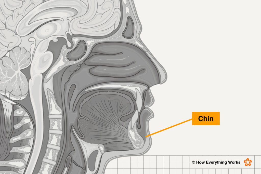
Humans are the only species to have evolved true chins, so the next time you rest yours thoughtfully in your hand, consider what an anatomical oddity you’re holding. Chimpanzees, our closest evolutionary relatives, don’t have chins. Neither do gorillas. Not even earlier humans, like Neanderthals, had a true chin. (A “true chin,” by the way, is defined as the bony bit at the center of your lower jaw that juts forward, not just any protuberance of flesh or fur, however adorable.)
Scientists have suggested that human chins evolved to strengthen the jaw during chewing, withstand the stresses of speech, or make us more appealing to potential mates (who doesn’t love a good chin?). But these theories aren’t perfect: The chin seems to be in the wrong place to reinforce the jaw, talking probably doesn’t produce enough force to require one, and chins aren’t limited to one sex. They may simply be a passive result of human faces becoming smaller and flatter as we evolved. For now, our chins remain uniquely ours — and uniquely puzzling.
The Vomeronasal Organ
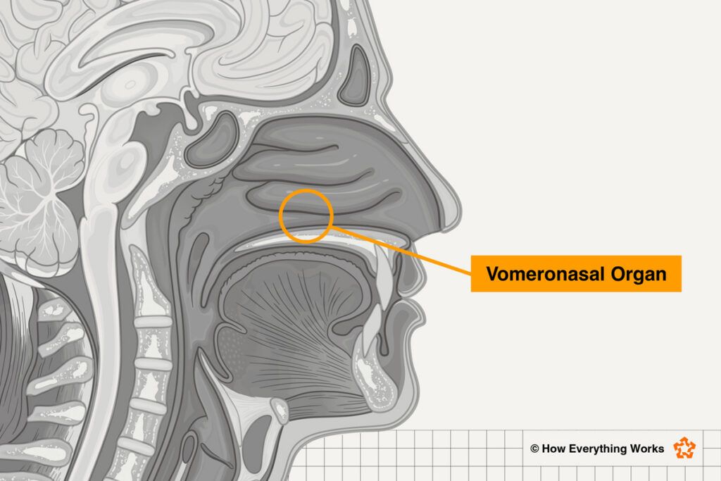
Numerous animals possess a vomeronasal organ, or VNO — a tiny structure in the nose that picks up chemical signals. Also called Jacobson’s organ, it helps influence social, sexual, and defensive behavior in many reptiles, amphibians, and mammals.
Whether humans have a working version has been debated for more than 300 years. Researchers have found tiny pits or ducts resembling the VNO in the nasal septum of some adults, and the structures may be more common in babies and children. But studies disagree considerably about how many people have them.
And simply finding the structure doesn’t prove that it works. In other animals, the VNO connects to specialized regions of the brain, but humans appear to be missing much of that equipment. Many of the genes used by other mammals to detect chemical signals through the VNO have also stopped working in humans. In other words, we lack both the hardware and the software to use the vomeronasal organ.
That has led some researchers to consider our VNO an evolutionary leftover rather than an active sense organ. Others aren’t ready to close the case. They point to possible traces of sensory tissue and rare tumors that have developed in the same area. For now, scientists can’t agree on whether the human VNO detects anything — much less whether it influences how we respond to other people, the way it does in other creatures.
The Placenta
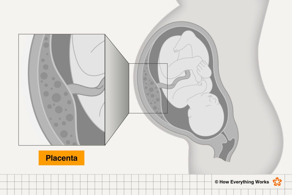
Every single human starts out dependent on the placenta, yet researchers have called it our least-understood organ. During pregnancy, this temporary body part grows from scratch and does much of the work that will eventually be taken over by the fetus’s lungs, kidneys, liver, digestive system, and immune system.
The placenta also acts as a go-between for two separate circulatory systems. Oxygen and nutrients pass from mother to fetus, while carbon dioxide and other waste travel in the opposite direction — all without their blood directly mixing. Meanwhile, the placenta produces hormones and reshapes blood vessels in the uterus to keep enough blood flowing through the system.
Part of the mystery comes down to a practical problem: Scientists need to see the placenta at work inside the womb, but they must do so without endangering parent or child. Much of what we know has therefore come from placentas examined after delivery. Researchers have compared this approach to trying to understand how a living body works by looking only at autopsy results. Animals aren’t always much help, either, since the human placenta differs significantly from those of many other mammals.
As a result, scientists still have major questions about how the placenta avoids attack by the parent’s immune system, why it works beautifully in some pregnancies but not in others, and how placental problems contribute to conditions such as preeclampsia and premature birth. They are also investigating how this short-lived organ might affect the health of both parent and child long after pregnancy ends.
The Appendix
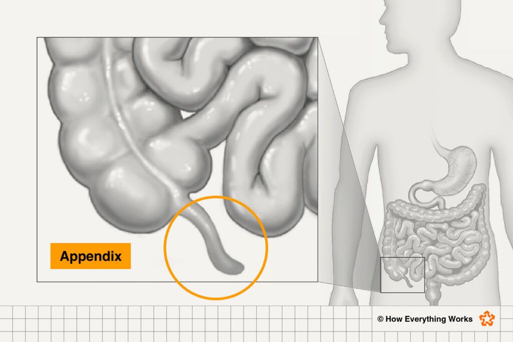
For generations, the appendix was the poster child for useless body parts: an evolutionary leftover that seemingly did nothing except become painfully inflamed and sometimes burst. Since people can live perfectly well after having it removed, there appeared to be little reason to think otherwise.
But the narrow pouch attached to the large intestine may not be merely taking up space. It contains plenty of immune tissue, suggesting that it helps the body recognize and respond to germs. Researchers have also proposed that the appendix serves as a safe house for beneficial gut bacteria. If an intestinal illness clears microbes out of the digestive tract, bacteria sheltered inside the appendix might emerge to help rebuild the gut’s microbial community.
The appendix’s evolutionary history makes its supposedly useless status even harder to defend. It has evolved independently numerous times among mammals and persisted for tens of millions of years. Animals with appendixes don’t share any obvious diet, habitat, or way of life, but their appendixes commonly contain immune tissue.
That doesn’t mean scientists have solved the mystery. They are still working out how much the appendix contributes to immunity and gut health, whether removing it has subtle long-term effects, and why evolution has apparently repeatedly favored a bacterial safe house with the unfortunate potential to send its owner to the emergency room.
The Interstitium
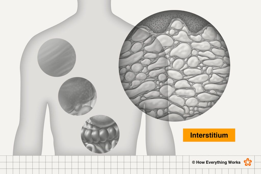
In 2018, researchers drew attention to a previously ignored network of fluid-filled spaces running through connective tissues around the body. Known as the interstitium, it can be found beneath the skin, around muscles, and in tissues lining the lungs, digestive tract, and urinary system.
Scientists already knew that fluid existed in the spaces between cells. What surprised them was how these particular spaces were organized — and how easily they had been hidden. To prepare tissue for traditional microscope slides, researchers drain away its fluids and treat it with preserving chemicals. That process caused the spaces to collapse, leaving behind what looked like a solid wall of collagen.
Researchers assumed that structure was unchanged from how it works inside us until they used an imaging technique that allowed them to examine living tissue without draining anything. When they did in the mid-2010s, they saw something quite different: open, interconnected spaces held up by bundles of collagen. One researcher compared the system to a fluid-filled highway.
Some scientists described the interstitium as a newly discovered organ, one that had somehow gone hidden until just a decade ago. But other scientists think that label goes too far. For now, it is safer to think of it as a widespread anatomical network that we are only beginning to understand.
The interstitium may cushion organs as they stretch and contract, help move cells and other materials through the body, and/or play a part in maintaining fluid balance. But researchers are still sorting out whether the interstitium truly acts as one connected system — or whether it is a collection of similar spaces with different jobs.
Hassall’s Corpuscles
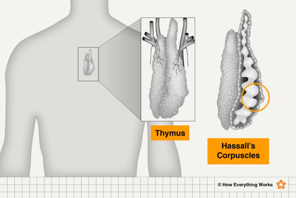
Inside the thymus, which is located behind the sternum and in front of the heart, are small, onion-shaped structures called Hassall’s corpuscles. British physician Arthur Hill Hassall first described them in the 1840s, so scientists have had nearly two centuries to study them. Even so, no one can yet give a complete account of what they do.
The thymus serves as a training center for T cells, important members of the immune system. There, young T cells learn how to recognize threats without attacking the body’s own tissues. Hassall’s corpuscles sit in the inner region of the thymus, right in the neighborhood where some of this crucial screening takes place.
Scientists once thought the corpuscles might simply be collections of aging cells or disposal sites for young T cells that flunked out of Thymus Academy. More recent research suggests that they take a much more active part in the process. Among other things, Hassall’s corpuscles release signals that affect nearby immune cells and may help produce regulatory T cells, which keep the immune system from turning against the body.
But these functions do not yet add up to a complete explanation. Hassall’s corpuscles differ considerably among species and are poorly represented in the mice commonly used for research. Recent three-dimensional imaging has found that they can occupy a substantial portion of the human thymic medulla — making it difficult to believe they are merely cellular leftovers, even if scientists still can’t say exactly what they do.